Under a microscope, the margin of a white clover (Trifolium repens) leaflet looks very different from the smooth green ‘clover leaf’ seen with the naked eye. Small teeth line the margin, and some cells around the teeth appear reddish-purple.
The previous version of this article listed several possible functions of serrations — increasing gas exchange through turbulence, increasing photosynthetic area, and discouraging herbivory — as though they were established for this white clover leaf. They were not.
This remaster separates what the images show from what developmental studies in other plants tell us about leaf margins.
What the images actually show
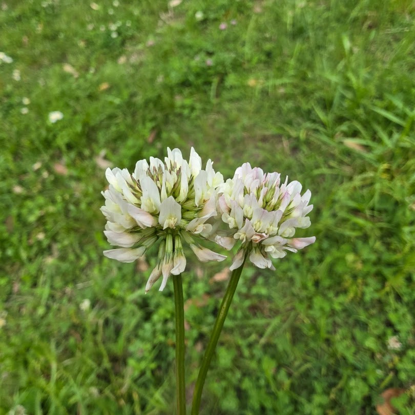
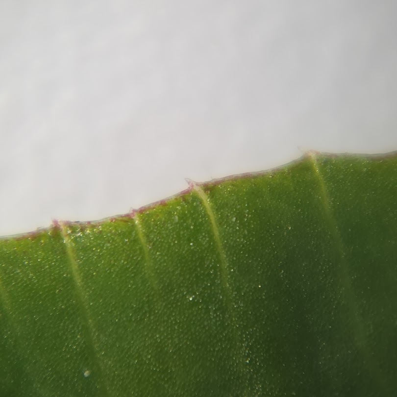
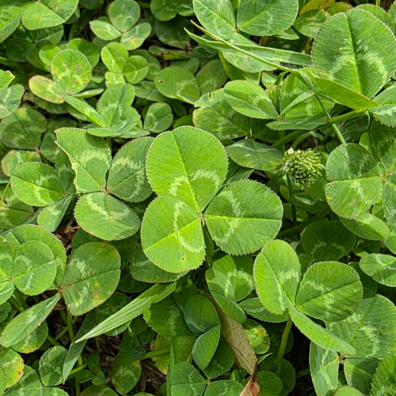
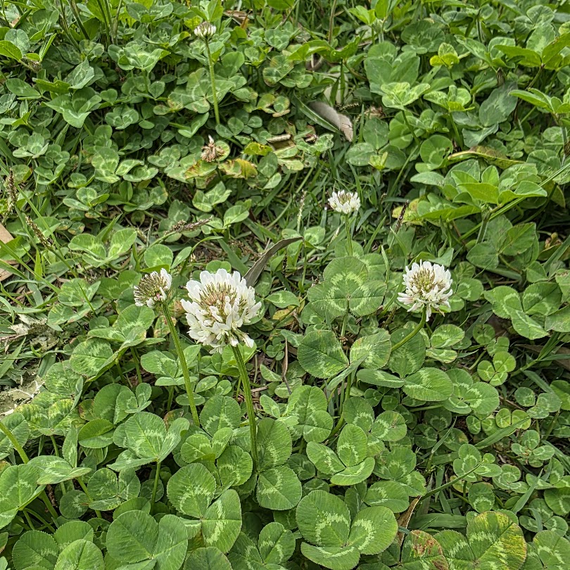
The photographs show repeated protrusions along the leaflet margin and cellular structure extending into the tooth region. The reliable observation is morphological: the margin is patterned by local differences in growth rather than being a continuous straight edge.
The reddish-purple cells are not chemically identified
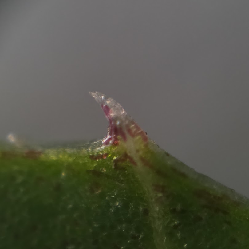
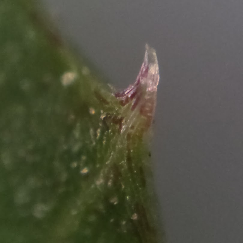
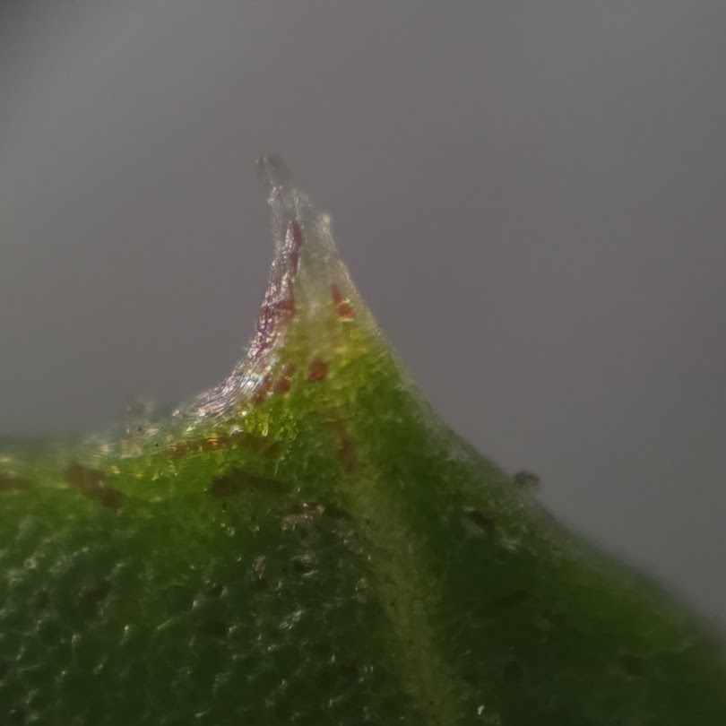
Some cells near the margin appear reddish-purple. Anthocyanins can produce similar colors in plants, but color in a photograph is not chemical identification. Confirming anthocyanins would require extraction, spectroscopy, chromatography, mass spectrometry, or another chemical assay.
The correct description is therefore that the cells appear to contain a reddish-purple pigment.
How are leaf teeth formed?
Leaf serration has been studied in detail in Arabidopsis. CUC transcription factors, PIN1-dependent auxin patterning, KNOX-related regulators and other developmental systems create spatial differences in growth along the margin. A 2025 review integrates genetic, hormonal, developmental and environmental regulation of leaf-margin shape.
Those mechanisms provide a useful framework, but the microscope images alone cannot establish which network controls these teeth in white clover.
Serrations do not have one universal function
Leaf-margin shape varies widely among species and can be associated with development, environment and ecological history. This observation does not demonstrate that white-clover teeth increase gas exchange, photosynthesis or herbivore resistance. Those claims have therefore been removed.
The more interesting point is developmental: a millimeter-scale tooth is the visible outcome of patterned cell growth, hormone distribution and gene regulation.
Why a simple microscope observation is still valuable
This is an owner observation, not a controlled experiment. Its value is that it creates better questions: Does the pigment change between young and mature leaves? Does light exposure change marginal coloration? Are tooth size and shape plastic? Can the pigment be chemically identified?
A familiar clover leaflet becomes a useful entry point into plant developmental biology once it is magnified.
For related context, see Purple Woodsorrel Petal Patterns Under the Microscope: Dark Veins Are Built from Differently Pigmented Cells.
For related context, see Why Do Leaf Midveins Taper? 147 Species Reveal Position-Specific Optimization for Transport and Mechanics.
For related context, see Upper and Lower Leaf Stomata Use Different K⁺ Channel Compositions for Light-Induced Opening.
References
- Kawamura E et al. Mechanisms of leaf tooth formation in Arabidopsis. The Plant Journal. 2010;62:429–441. https://doi.org/10.1111/j.1365-313X.2010.04156.x
- Lu Z et al. A Review of Regulation of Leaf Margin Development: Integrating Genetic, Hormonal, and Environmental Controls. Physiologia Plantarum. 2025;177:e70627. https://doi.org/10.1111/ppl.70627

Comments