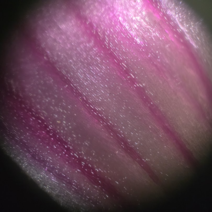
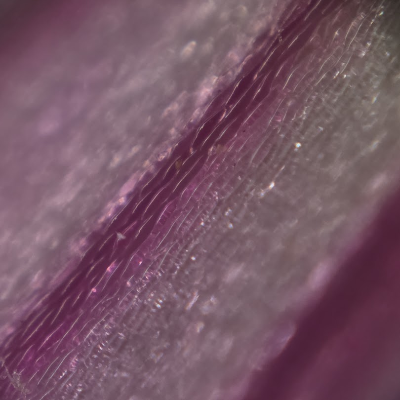
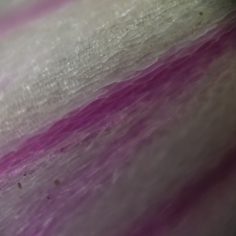
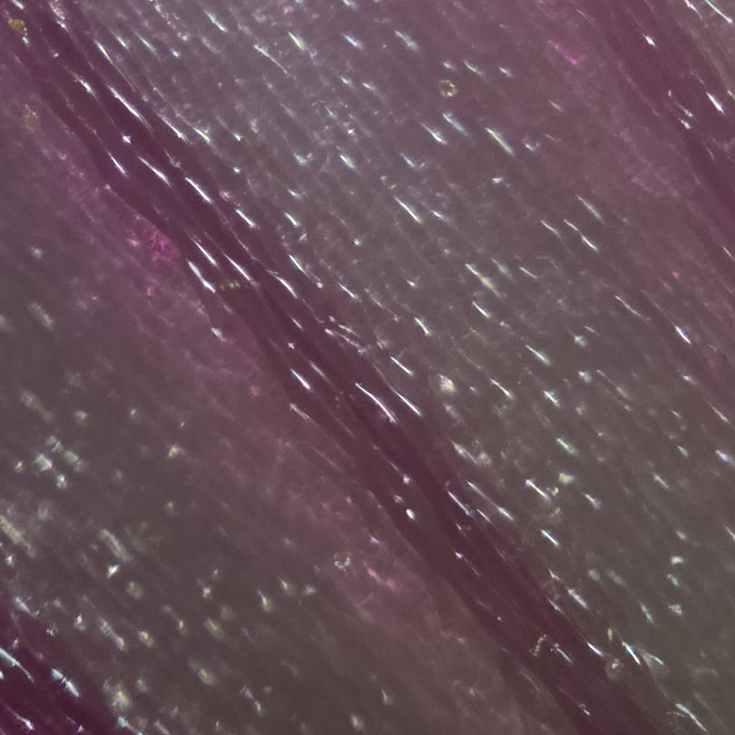
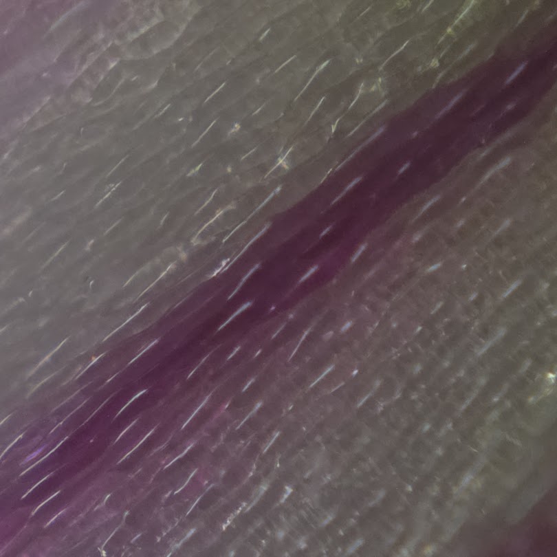
To the naked eye, a purple woodsorrel flower appears to have dark purple veins painted across a pale pink-purple petal. Under magnification, that continuous pattern resolves into groups of cells with different pigmentation.
This is an owner observation rather than a controlled experiment, so it is useful to separate what the images directly show from what flower-pigment biology allows us to infer.
- The dark petal veins resolve into cellular regions
- Higher magnification reveals darker and lighter cells side by side
- The photographs do not chemically identify anthocyanins
- Cell-by-cell pigmentation reflects spatial developmental regulation
- A related woodsorrel article concerns a different phenomenon
- References
The dark petal veins resolve into cellular regions
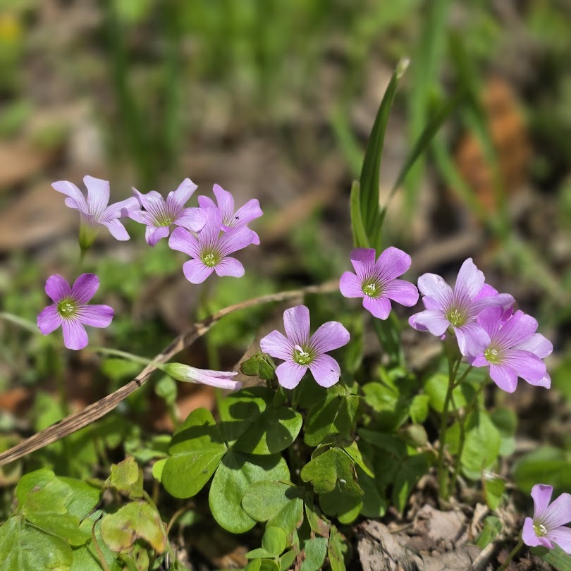
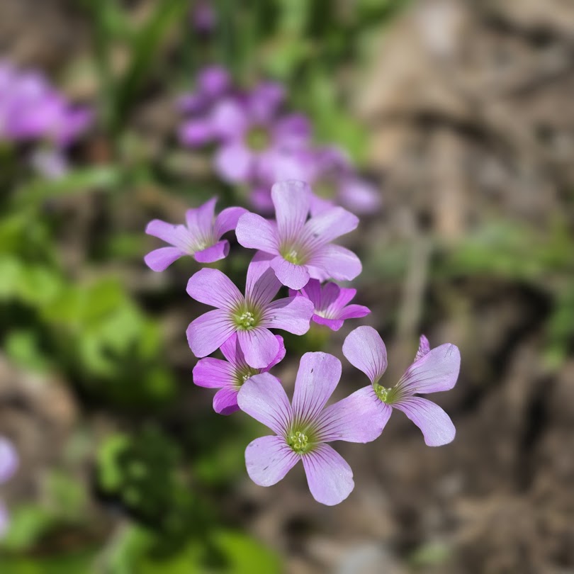
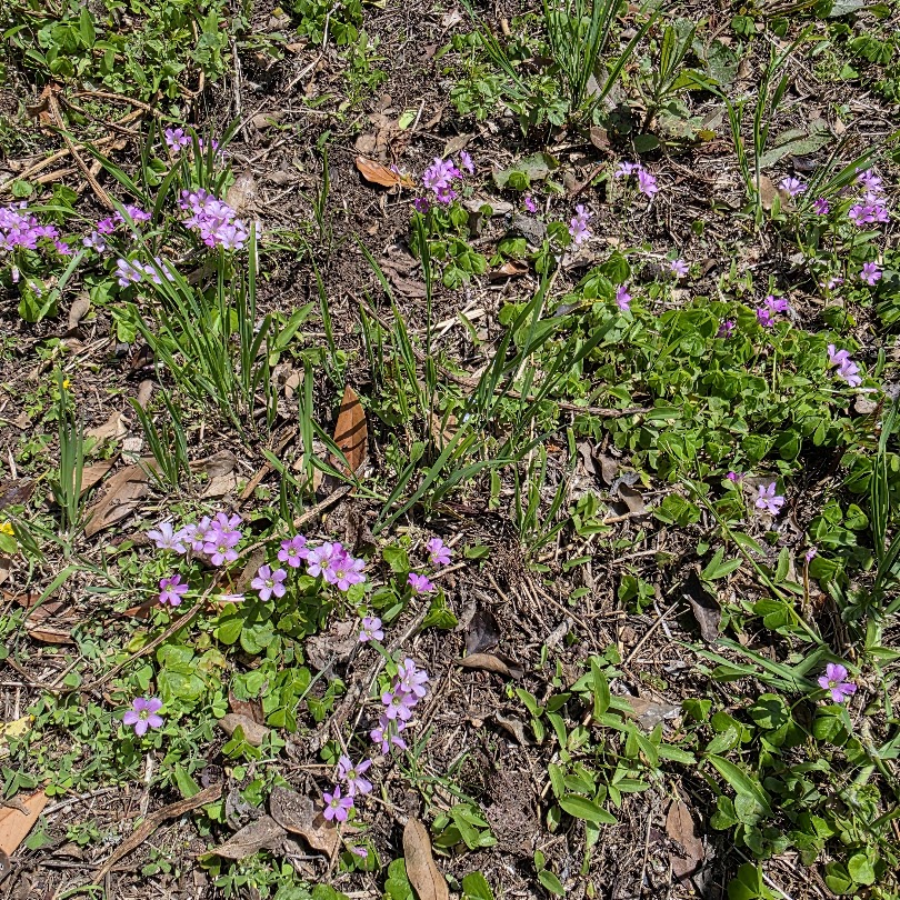
At low magnification, the dark lines are not a separate painted structure. Darker regions follow the arrangement of petal cells, while paler cell groups occupy the surrounding tissue. The macroscopic pattern is therefore produced by spatial differences in pigmentation across cells.
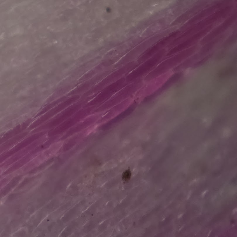
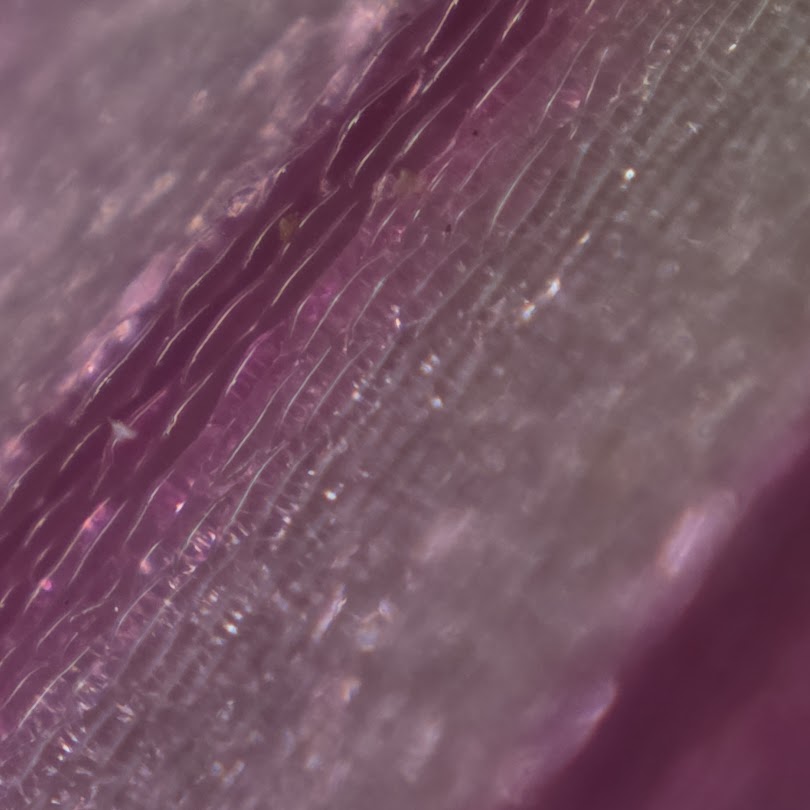
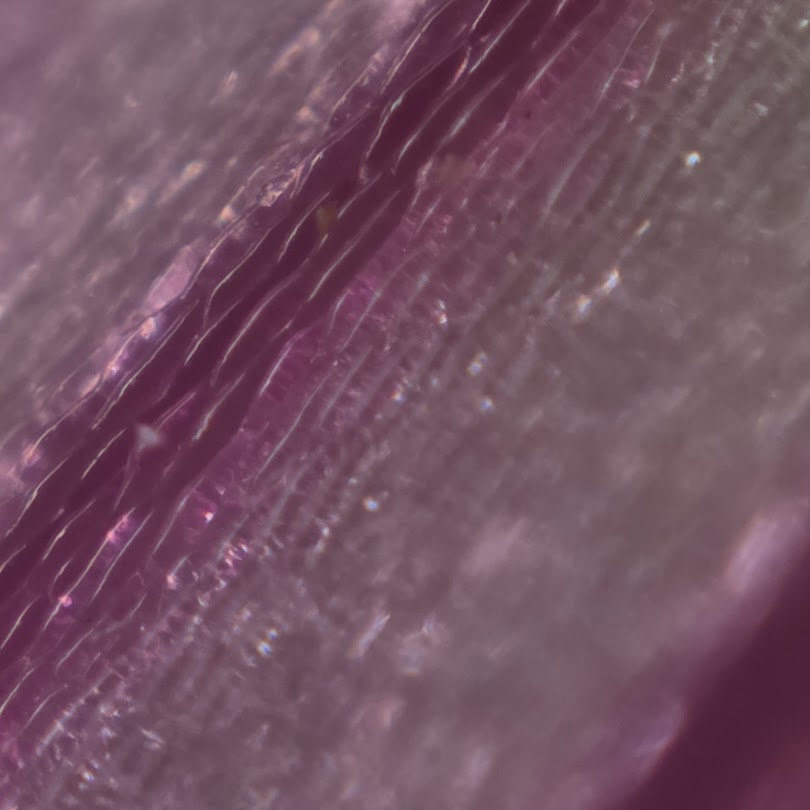
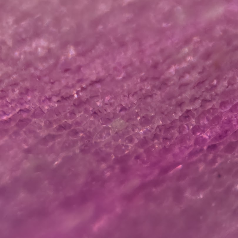
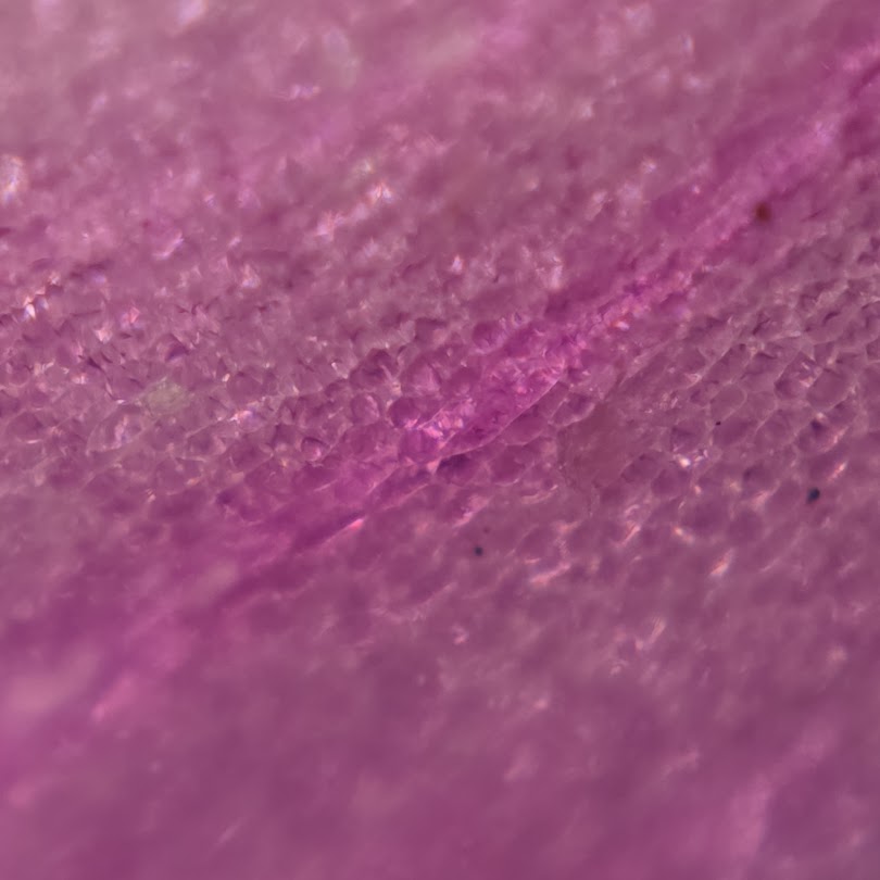
Higher magnification reveals darker and lighter cells side by side
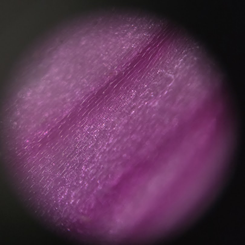
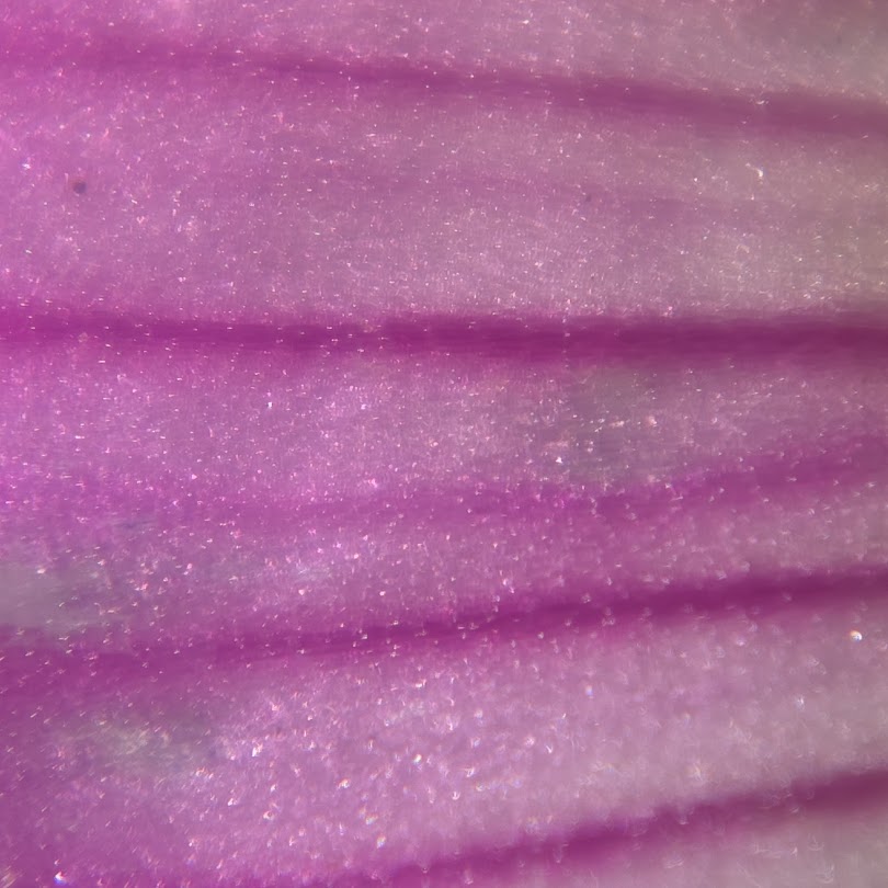
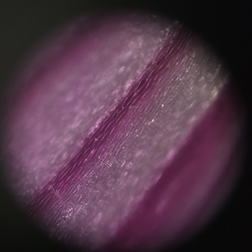
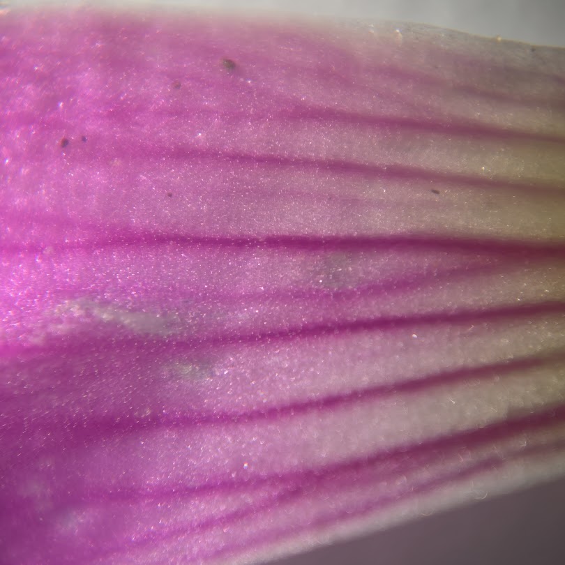
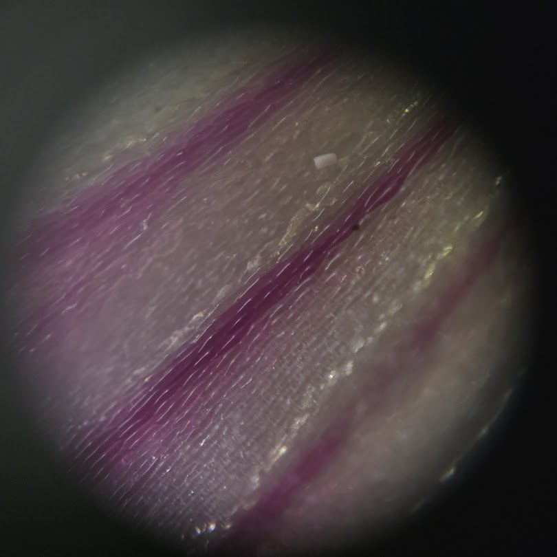
At higher magnification, individual epidermal-cell outlines become clearer. Cells in the dark-vein region appear strongly reddish-purple, whereas neighboring regions contain paler cells. Some transitions look gradual, but the visible petal pattern can be decomposed into a mosaic of differently pigmented cells.
The photographs do not chemically identify anthocyanins
Anthocyanins commonly produce red, purple and blue flower colors and are usually stored in vacuoles. Flower color can also vary with anthocyanin identity and concentration, vacuolar pH, copigments and cell structure.
However, photographs alone cannot identify which pigments are present in this woodsorrel. Chemical confirmation would require extraction and analytical methods such as spectroscopy, chromatography or mass spectrometry.
Cell-by-cell pigmentation reflects spatial developmental regulation
Flower patterns can arise when pigment-biosynthesis genes and transcription factors are active in different spatial domains. What appears microscopically as a dark or pale cell is the downstream result of how that cell synthesized, transported and stored pigments.
This observation does not identify the patterning genes in purple woodsorrel, but it clearly shows how a whole-petal pattern resolves to cellular-scale differences.
A related woodsorrel article concerns a different phenomenon
An earlier ChloroQuest article examined green-versus-red leaf polymorphism and urban heat adaptation in Oxalis corniculata. The present observation concerns petal pigmentation in purple woodsorrel, so species, organ and biological question are different.
Once the full archive remaster is complete, these articles can be connected as separate layers of an Oxalis color cluster: leaf color, flower color, environmental adaptation and microscopy.
For related context, see Why Are Urban Oxalis Leaves Red? Urban Heat Islands Favor a Heat-Tolerant Leaf-Color Phenotype.
For related context, see Microscope View of White Clover Leaf Teeth — What Can We Really Infer from the Reddish-Purple Margin?.
For related context, see How miR159 and CKX6 Control Rose Petal Size Through Cytokinin Turnover.
For related context, see Eustoma Cultivation Log, 2023: From Seedling Arrival to 2.5 Months After Potting.
References
- Grotewold E. The genetics and biochemistry of floral pigments. Annual Review of Plant Biology. 2006;57:761–780.
- Plant anthocyanins: classification, biosynthesis, regulation, bioactivity, and health benefits. Plant Physiology and Biochemistry. 2024;217:109268. https://doi.org/10.1016/j.plaphy.2024.109268

Comments